Body Fluids and Circulation – Regulation of Cardiac Activity: NEET MCQs with Answers
9 free questions on Body Fluids and Circulation – Regulation of Cardiac Activity for NEET (UG), starting with previous-year NEET and AIPMT questions. Try each one, then tap Show answer for the answer and a short explanation.
Syllabus covered: Regulation of Cardiac Activity
Question 1
Previous-year questionWhich of the following regulate the cardiac activities of the heart? [NEET 2025]
A. Nodal tissue
B. A special neural centre in the medulla oblongata
C. Adrenal medullary hormones
D. Adrenal cortical hormones
A. Nodal tissue
B. A special neural centre in the medulla oblongata
C. Adrenal medullary hormones
D. Adrenal cortical hormones
- A, B and C only
- A, B, C and D
- A, C and D only
- A, B and D only
Show answer
Answer: (A) A, B and C only
Explanation: Normal cardiac activity is auto-regulated by the nodal tissue (A), and can be moderated by a special neural centre in the medulla oblongata through autonomic nerves (B) and by adrenal medullary hormones that increase cardiac output (C). Adrenal cortical hormones are not listed in NCERT as regulators of cardiac activity.
Question 2
Previous-year questionWhich one of the components of a normal human ECG is correctly interpreted? [2011]
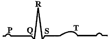
- Peak P and peak R together — systolic and diastolic blood pressures
- Complex QRS — one complete pulse
- Peak P — initiation of left atrial contraction only
- Peak T — initiation of total cardiac contraction
Show answer
Answer: (B) Complex QRS — one complete pulse
Explanation: Each QRS complex marks one ventricular depolarisation and therefore one heart beat, so counting the complexes over a minute gives the pulse rate. The P wave initiates contraction of both atria and not the left alone, the T wave marks ventricular repolarisation, and the ECG records electrical activity rather than blood pressure.
Question 3
Previous-year questionIn a standard ECG, which of the following is the correct representation of the corresponding activity of the human heart? [2009]
- P — depolarisation of the atria
- S — start of systole
- R — repolarisation of the ventricles
- T — end of diastole
Show answer
Answer: (A) P — depolarisation of the atria
Explanation: The P wave records the spread of depolarisation over the atria, which immediately precedes atrial contraction. The QRS complex represents ventricular depolarisation and the T wave ventricular repolarisation, so the descriptions in the other options are misassigned.
Question 4
Previous-year questionThe bundle of His is a part of which organ in humans? [2010]
- Kidney
- Brain
- Pancreas
- Heart
Show answer
Answer: (D) Heart
Explanation: It forms part of the conducting system of the heart, carrying the impulse from the atrioventricular node to the ventricular musculature and so ensuring that the ventricles contract from the apex upward, a moment after the atria.
Question 5
Previous-year questionThe bundle of His is a network of [2004]
- muscle fibres found only in the ventricle wall
- nerve fibres found throughout the heart
- nerve fibres distributed in the ventricles
- muscle fibres distributed throughout the heart walls
Show answer
Answer: (A) muscle fibres found only in the ventricle wall
Explanation: The bundle of His consists of modified cardiac muscle fibres, not nerve fibres, and it runs from the atrioventricular node into the interventricular septum, dividing into right and left branches that supply the two ventricles. Its specialisation is for rapid conduction rather than contraction.
Question 6
Previous-year questionThe cardiac pacemaker of a patient fails to function normally and the doctors decide to graft an artificial pacemaker. It is likely to be grafted at the site of the [2003]
- sinuatrial node
- atrioventricular bundle
- atrioventricular node
- Purkinje system
Show answer
Answer: (A) sinuatrial node
Explanation: Since the natural rhythm is set by the sinuatrial node, an artificial pacemaker is placed so as to take over its function, delivering regular electrical impulses to the right atrium from which they spread through the normal conducting pathway.
Question 7
Previous-year questionThe correct route through which the pulse-making impulse travels in the heart is [1995]
- SA node → Purkinje fibres → bundle of His → AV node → heart muscles
- AV node → bundle of His → SA node → Purkinje fibres → heart muscles
- SA node → AV node → bundle of His → Purkinje fibres → heart muscles
- AV node → SA node → Purkinje fibres → bundle of His → heart muscles
Show answer
Answer: (C) SA node → AV node → bundle of His → Purkinje fibres → heart muscles
Explanation: The impulse originates in the sinuatrial node, spreads through the atrial musculature to the atrioventricular node, passes down the bundle of His into its two branches within the interventricular septum, and is finally distributed to the ventricular muscle by the Purkinje fibres.
Doing well? Create a free account to take timed tests on this topic and see your weak areas.
Question 8
Previous-year questionContraction of the ventricle in the heart begins with the command from the [1999]
- Purkinje fibres
- chordae tendinae
- AV node
- SA node
Show answer
Answer: (D) SA node
Explanation: Every beat, including ventricular systole, is initiated by the sinuatrial node; the impulse spreads over the atria to the atrioventricular node, which delays it briefly and then relays it through the bundle of His and the Purkinje fibres to the ventricular muscle.
Question 9
Previous-year questionThe impulse of the heart beat originates from the [2001]
- vagus nerve
- SA node
- cardiac nerve
- AV node
Show answer
Answer: (B) SA node
Explanation: The beat arises within the heart itself, at the sinuatrial node. The vagus and the sympathetic cardiac nerves only modify the rate that the node sets, the vagus slowing it and the sympathetic supply accelerating it.
Want more Body Fluids and Circulation – Regulation of Cardiac Activity questions?
E-GYAN NIDHI has 93 questions on this topic and 63,000+ in total, all free: timed CBT tests, NEET marking and instant analysis.
Register free Free Sunday Mock TestMore topics in Human Physiology
- Respiration – Respiratory Volumes and Capacities MCQs
- Body Fluids and Circulation – Blood MCQs
- Excretion – Modes of Excretion MCQs
- Locomotion and Movement – Disorders of muscular and skeletal system-Myasthenia gravis MCQs
- Neural Control and Coordination – Central Neural System MCQs
- Endocrine System – Chemical Coordination and Integration MCQs
- Respiration – Human Respiratory System MCQs
- Respiration – Mechanism of Breathing its regulation in humans MCQs
- Respiration – Exchange of Gases MCQs
- Respiration – Transport of Gases and Regulation of Respiration MCQs
- Respiration – Disorders of Respiratory System MCQs
- Body Fluids and Circulation – Circulatory Pathways MCQs
- Body Fluids and Circulation – Disorders of Circulatory System MCQs
- Excretion – Human Excretory System MCQs
- Excretion – Urine Formation MCQs
- Excretion – Function of Tubules MCQs
- Excretion – Mechanism of Concentration of the Filtrate MCQs
- Excretion – Regulation of Kidney Function MCQs
- Excretion – Disorders of Excretory System MCQs
- Locomotion and Movement – Joints MCQs
- Locomotion and Movement – Skeletal System and its functions (To be dealt with the MCQs
- Locomotion and Movement – Skeletal Muscle and Its Contraction MCQs
- Endocrine System – Mechanism of Hormone Action (Elementary Idea) MCQs
- Endocrine System – Hypo-and hyperactivity and related disorders (Common MCQs
- Neural Control and Coordination – Neuron as Structural and Functional Unit of Neural System MCQs
- Neural Control and Coordination – Human Neural System MCQs