Human Reproduction – Female Reproductive System: NEET MCQs with Answers
15 free questions on Human Reproduction – Female Reproductive System for NEET (UG), starting with previous-year NEET and AIPMT questions. Try each one, then tap Show answer for the answer and a short explanation.
Syllabus covered: Female Reproductive System: Microscopic anatomy of ovary
Question 1
Previous-year questionThe figure given below depicts a diagrammatic sectional view of the female reproductive system of humans. Which one set of three parts out of A–F have been correctly identified? [AIPMT 2011]
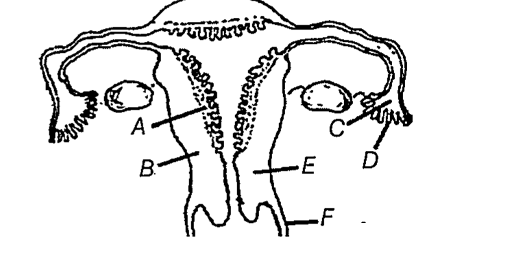
- C–Infundibulum, D–Fimbriae, E–Cervix
- D–Oviducal funnel, E–Uterus, F–Cervix
- A–Perimetrium, B–Myometrium, C–Fallopian tube
- B–Endometrium, C–Infundibulum, D–Fimbriae
Show answer
Answer: (A) C–Infundibulum, D–Fimbriae, E–Cervix
Explanation: The Fallopian tube is about 10–12 cm long and extends from the periphery of each ovary to the uterus. Its part closest to the ovary is the funnel-shaped infundibulum (C), whose edges bear finger-like fimbriae (D) that collect the ovum after ovulation; the uterus opens into the vagina through the narrow cervix (E).
Question 2
Previous-year questionBartholin's glands are situated [AIPMT 2003]
- on either side of vagina in humans
- on either side of vas deferens in humans
- on the sides of the head of some amphibians
- at the reduced tail end of birds
Show answer
Answer: (A) on either side of vagina in humans
Explanation: Bartholin's glands (bulbovestibular glands) are a pair of small glands lying just behind the labia minora, one on either side of the vaginal orifice. They secrete mucus that lubricates the vagina during coitus and parturition.
Question 3
Previous-year questionThe part of Fallopian tube closest to the ovary is [AIPMT 2010]
- isthmus
- infundibulum
- cervix
- ampulla
Show answer
Answer: (B) infundibulum
Explanation: The infundibulum is the funnel-shaped proximal part of the oviduct nearest the ovary. Its margin bears fimbriae which help in collecting the ovum released at ovulation.
Question 4
Previous-year questionStatement I: Vas deferens receives a duct from seminal vesicle and opens into urethra as the ejaculatory duct. [NEET 2023] Statement II: The cavity of the cervix is called cervical canal, which along with vagina forms birth canal. In the light of the above statements, choose the correct answer from the options given below.
- Both statement I and statement II are false
- Statement I is true, but statement II is false
- Statement I is false, but statement II is true
- Both statement I and statement II are true
Show answer
Answer: (D) Both statement I and statement II are true
Explanation: Both statements are correct. The vas deferens is joined by the duct of the seminal vesicle to form the ejaculatory duct, which opens into the urethra. The cavity of the cervix is the cervical canal, which together with the vagina forms the birth canal.
Question 5
Previous-year questionGiven below are two statements. [NEET 2024] Statement I: The presence or absence of hymen is not a reliable indicator of virginity. Statement II: The hymen is torn during the first coitus only. In the light of the above statements, choose the correct answer from the options given below.
- Both Statement I and Statement II are true
- Both Statement I and Statement II are false
- Statement I is true, but Statement II is false
- Statement I is false, but Statement II is true
Show answer
Answer: (C) Statement I is true, but Statement II is false
Explanation: Statement I is true — the hymen is not a reliable indicator of virginity. Statement II is false because the hymen may be torn by a sudden fall or a jolt during horse-riding, cycling, gymnastics or dancing, and in some women it persists even after coitus.
Question 6
Previous-year questionWhich of the following is not a component of Fallopian tube? [NEET 2024]
- Uterine fundus
- Isthmus
- Infundibulum
- Ampulla
Show answer
Answer: (A) Uterine fundus
Explanation: The Fallopian tube consists of the funnel-shaped infundibulum with fimbriae, the wide ampulla and the narrow isthmus that joins the uterus. The fundus is the dome-shaped upper part of the uterus itself, not a part of the Fallopian tube.
Question 7
Previous-year questionAfter ovulation the collapsed ovarian follicle shrinks and becomes filled with cells to form the [2003]
- corpus atresia
- corpus luteum
- corpus adiposum
- corpus albicans
Show answer
Answer: (B) corpus luteum
Explanation: After ovulation the remaining cells of the ruptured Graafian follicle transform into the corpus luteum, which secretes large amounts of progesterone. If fertilisation does not occur, the corpus luteum degenerates into the corpus albicans.
Doing well? Create a free account to take timed tests on this topic and see your weak areas.
Question 8
Previous-year questionThe part of the fallopian tube closest to the ovary is the [2010]
- cervix
- isthmus
- ampulla
- infundibulum
Show answer
Answer: (D) infundibulum
Explanation: The funnel-shaped infundibulum, with its fringe of finger-like fimbriae, lies nearest the ovary and collects the released ovum. It leads into the wide ampulla, where fertilisation normally occurs, and thence through the narrow isthmus to the uterus.
Question 9
Previous-year questionThe oestrous cycle is characteristic of [1995]
- mammalian males other than primates
- human males only
- mammalian females other than primates
- human females only
Show answer
Answer: (C) mammalian females other than primates
Explanation: Non-primate mammals such as the cow, dog and rat show an oestrous cycle, in which the female is receptive only during oestrus and the endometrium is reabsorbed rather than shed if fertilisation fails. Primates, including humans, show a menstrual cycle instead, with cyclic shedding of the endometrium.
Question 10
Which sequence correctly lists the parts of the human female reproductive tract, starting at the ovary and ending at the opening to the exterior?
- Ovary → uterus → isthmus → ampulla → cervical canal → vagina
- Ovary → isthmus → ampulla → infundibulum → uterus → cervical canal → vagina
- Ovary → infundibulum → ampulla → isthmus → uterus → cervical canal → vagina
- Ovary → infundibulum → ampulla → isthmus → cervical canal → uterus → vagina
Show answer
Answer: (C) Ovary → infundibulum → ampulla → isthmus → uterus → cervical canal → vagina
Explanation: The funnel-shaped infundibulum lies closest to the ovary and leads into the wider ampulla, followed by the narrow isthmus, which joins the uterus. The uterus opens into the vagina through the cervical canal. The cervical canal and vagina together form the birth canal.
Question 11
Which one of the following is NOT a part of the female external genitalia?
- Vagina
- Labia minora
- Mons pubis
- Hymen
Show answer
Answer: (A) Vagina
Explanation: The female external genitalia are the mons pubis, labia majora, labia minora, hymen and clitoris. The vagina is an internal muscular tube that serves as a female accessory duct and part of the birth canal.
Question 12
The last part of the oviduct, which has a narrow lumen and joins the uterus, is the
- infundibulum
- isthmus
- fimbriae
- ampulla
Show answer
Answer: (B) isthmus
Explanation: Going from the ovary towards the uterus, the oviduct has the funnel-shaped infundibulum bearing fimbriae, then the wider ampulla, and finally the isthmus. The isthmus has a narrow lumen and joins the uterus.
Question 13
Which of the following is a function of the human ovary?
- Secreting milk after childbirth
- Providing the site of fertilisation
- Producing the female gamete and steroid hormones
- Housing the developing foetus
Show answer
Answer: (C) Producing the female gamete and steroid hormones
Explanation: The ovaries are the primary female sex organs: they produce the ovum and the ovarian steroid hormones, estrogen and progesterone. Fertilisation takes place in the oviduct, the foetus develops in the uterus, and milk is made by the mammary glands.
Question 14
The circular, darkly pigmented area of skin that surrounds the nipple of the breast is called the
- mammary ampulla
- alveolus
- lactiferous duct
- areola
Show answer
Answer: (D) areola
Explanation: The pigmented ring of skin around the nipple is the areola. Alveoli are milk-secreting cell clusters inside the gland, while the mammary ampulla and lactiferous duct are internal channels that carry milk.
Question 15
In the mammary gland of a lactating woman, milk is secreted by
- the cells of the alveoli
- the lining of the lactiferous duct
- the mammary ampulla
- the fat cells of the breast
Show answer
Answer: (A) the cells of the alveoli
Explanation: The glandular tissue of each breast forms lobes containing clusters of cells called alveoli. These alveolar cells secrete milk, including the colostrum of the first few days, which is stored in their lumens. Tubules, ducts, the ampulla and the lactiferous duct only carry the milk to the nipple.
Want more Human Reproduction – Female Reproductive System questions?
E-GYAN NIDHI has 67 questions on this topic and 63,000+ in total, all free: timed CBT tests, NEET marking and instant analysis.
Register free Free Sunday Mock TestMore topics in Reproduction
- Human Reproduction – Male Reproductive System MCQs
- Human Reproduction – Gametogenesis-spermatogenesis & oogenesis MCQs
- Human Reproduction – Menstrual Cycle MCQs
- Human Reproduction – Fertilisation and Implantation MCQs
- Human Reproduction – Pregnancy and Embryonic Development MCQs
- Human Reproduction – Parturition and Lactation MCQs
- Human Reproductive Health – Population Stabilisation and Birth Control MCQs
- Human Reproductive Health – Amniocentesis MCQs
- Human Reproductive Health – Sexually Transmitted Infections (STIs) MCQs
- Human Reproductive Health – Infertility MCQs